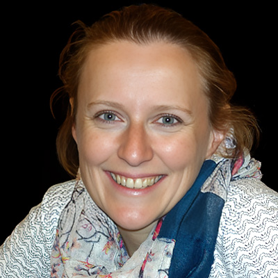
Marine Laporte
Univ-Lyon, France
Education
2011-15 – PhD thesis, Pr. Sadoul lab, Institut des Neurosciences, Grenoble, FR
2010-11 – Master 2 in Neurosciences (rank 3/32), Grenoble Alpes University, Grenoble, FR
2009-10 – Master 1 in Cell Biology (rank 4/56), Grenoble Alpes University, Grenoble, FR
2006-09- Bachelor’s in biology (rank 16/186), Grenoble Alpes University, Grenoble, FR
Professional career (in reverse chronological order)
• 2023 – Full researcher (CRCN INSERM) – Durand lab – University of Claude Bernard Lyon 1 – France
Study of membrane remodeling during cilia and flagella formation
1 publication
People management: Master 2 (2024 – 6 month), Master 1 (2024 – 3 month, 2024 – 3 month, 2023, 2
month)
• 2019-2023 post-doctoral fellow – Guichard & Hamel lab – University of Geneva – Switzerland
Investigation of centriolar assembly and function of structural centriolar elements.
9 publications – 2 reviews
People management: Ph.D student (2021 – 12 month), Technician (2020 – 6 month), Master 2 (2020-
21 – 18 month), Master 1 ( 2019 – 3 month), High school / middle school (2019, 2021 – 1 week).
• 2016-2018 post-doctoral fellow – Martinou lab – University of Geneva – Switzerland
Role of mitochondrial metabolism in neuroal excitability.
2 publications
People management: BTS (2017 – 3 month), Technician student (2017 – 12 month)
• 2011-2015 Ph.D thesis – Sadoul lab – University of Grenoble Alpes – France Function of Alix in
neuronal development and maturation.
4 publications – 1 review
People management: Master 2 (2014 – 6 month; 2015 – 6 month), Master 1 (2013 – 3 month),
Bachelor 3 (2013 – 1 month), High school / middle school (2012, 2013, 2014 – 1 week).
Visualizing the native cellular organization by coupling cryo-fixation with expansion microscopy (Cryo-ExM)
Abstract
Super-resolution fluorescent microscopy (SRM), encompassing expansion microscopy (ExM) since few years now, allows to locate proteins with nanometer resolution in a cellular context. However, SRM often requires cell fixation with aldehyde-based chemical crosslinkers, such as paraformaldehyde, or protein precipitation with cold methanol which potentially alter the native cellular state and the following interpretations. Cryo-fixation has proven to be the gold standard for efficient preservation of the native cell ultrastructure compared to chemical fixation, however it is not widely used in fluorescence microscopy owing to implementation. We recently developed a method combining cryo-fixation to ExM (Cryo-ExM), which allow nanoscale observation of a wide cellular compartment in their native state. We could demonstrate that Cryo-ExM allows the native preservation of membrane-based organelles such as mitochondria, endoplasmic reticulum, golgi and lysosomes together with the cytoskeleton component actin and microtubules and preserve all the structure in the same way, contrary to chemical fixations. Moreover, direct comparison with the gold-standard chemical fixation PFA-GA for the preservation of cellular structure, demonstrate that cryo-fixation bypassed drawbacks associated with this chemical fixation such as antigen accessibility due to strong protein-protein crosslinking. Overall.
In summary, we introduce a new method to perform super-resolution expansion microscopy by coupling cryo-fixation of a biological specimen with ExM, providing a universal framework to visualize subcellular compartments without chemical fixation artefacts. Importantly, this method also demonstrates that the classical cryo-substitution protocols developed for electron microscopy are compatible with expansion microscopy by replacing the EM resin with hydrogel monomer solutions. Therefore, this approach may also be applicable on tissues cryo-fixed by high-pressure freezing as well as in hydrogel-based tissue clearing. Finally, as expansion microscopy is also compatible with SIM, STED or dSTORM 18–20, our method now allows all these microscopy modalities to image cells in their native state, paving the way for further studies of complex and rapid dynamic cellular processes.